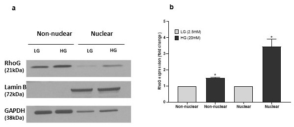
Fig. 3. Chronic exposure to high glucose promotes nuclear association of RhoG in INS-1 832/13 cells. Panel a: INS-1 832/13 cells were exposed to either basal glucose (LG, 2.5 mM) or high glucose (HG, 20 mM) for 24 hours. Following incubation, total nuclear and cytosolic fractions were isolated, and relative abundance of RhoG was determined in these fractions by Western blotting. GAPDH and Lamin B were used as marker proteins for non-nuclear and nuclear fractions, respectively. A representative blot from four independent studies is shown here. Panel b: Densitometric quantitation of relative abundance of RhoG in cytosolic and nuclear fractions isolated form basal and high glucose treated INS-1 832/13 cells is shown here. The data are expressed as fold change relative to respective LG (mean ± SEM; n=4 ; * p< 0.05).